


Clavicle fracture is one of the most common fractures, accounting for 2.6%-4% of all fractures. Due to the anatomical characteristics of the midshaft of the clavicle, midshaft fractures are more common, accounting for 69% of clavicle fractures, while fractures of the lateral and medial ends of the clavicle account for 28% and 3% respectively.
As a relatively uncommon type of fracture, unlike midshaft clavicle fractures that are caused by direct shoulder trauma or force transmission from upper limb weight-bearing injuries, fractures of the medial end of the clavicle are commonly associated with multiple injuries. In the past, the treatment approach for fractures of the medial end of the clavicle has typically been conservative. However, studies have shown that 14% of patients with displaced fractures of the medial end may experience symptomatic nonunion. Therefore, in recent years, more and more scholars have leaned towards surgical treatment for displaced fractures of the medial end that involve the sternoclavicular joint. However, the medial clavicular fragments are usually small, and there are limitations to fixation using plates and screws. Local stress concentration remains a challenging issue for orthopedic surgeons in terms of effectively stabilizing the fracture and avoiding fixation failure.

I.Distal Clavicle LCP Inversion
The distal end of the clavicle shares similar anatomical structures with the proximal end, both having a broad base. The distal end of the clavicle locking compression plate (LCP) is equipped with multiple locking screw holes, allowing for effective fixation of the distal fragment.
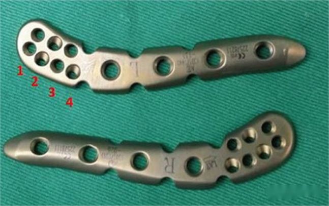
Taking into account the structural similarity between the two, some scholars have placed a steel plate horizontally at a 180° angle at the distal end of the clavicle. They have also shortened the part originally used to stabilize the distal end of the clavicle and found that the internal implant fits snugly without the need for shaping.

Placing the clavicle’s distal end in an inverted position and fixing it with a bone plate on the medial side has been found to provide satisfactory fit.


In a case of a 40-year-old male patient with a fracture at the medial end of the right clavicle, an inverted distal clavicle steel plate was used. A follow-up examination 12 months after the surgery indicated a good healing outcome.
Inverted distal clavicle locking compression plate (LCP) is a commonly used internal fixation method in clinical practice. The advantage of this method is that the medial bone fragment is held by multiple screws, providing a more secure fixation. However, this fixation technique requires a sufficiently large medial bone fragment for optimal results. If the bone fragment is small or there is intra-articular comminution, the fixation effectiveness may be compromised.
II. Dual Plate Vertical Fixation Technique
The dual plate technique is a commonly used method for complex comminuted fractures, such as fractures of the distal humerus, comminuted fractures of the radius and ulna, and so on. When effective fixation cannot be achieved in a single plane, dual locking steel plates are used for vertical fixation, creating a dual-plane stable structure. Biomechanically, dual plate fixation offers mechanical advantages over single plate fixation.

The upper fixation plate

The lower fixation plate and four combinations of dual plate configurations


Post time: Jun-12-2023


