


The selection of the entry point for Intramedullary of Tibial Fractures is one of the key steps in the success of surgical treatment. A poor entry point for Intramedullary, whether in the suprapatellar or infrapatellar approach, may result in loss of repositioning, angular deformity of the fracture end, and injury to the vital structures of the knee around the entry point.
The 3 aspects of the tibial intramedullary nail insertion point will be described.
What is the standard tibial intramedullary nail insertion point?
What are the effects of a deviated tibial intramedullary nail?
How is the correct point of entry determined intraoperatively?
I. What is the standard point of entry for Tibial Intramedullary?
The orthotopic position is located at the intersection of the mechanical axis of the tibia and the tibial plateau, the medial edge of the lateral intercondylar spine of the tibia, and the lateral position is located on the watershed between the tibial plateau and the tibial stem migration zone.
Range of safety zone at the entry point
22.9±8.9mm, in which area the needle can be inserted without damaging the bony stop of the ACL and the meniscus tissue.

II. What are the effects of a deviated Tibial Intramedullary Nail?
Depending on the proximal, middle, and Distal Tibial Fractures, the proximal tibial fracture has the most pronounced effect, the middle tibial fracture has the least effect, and the distal end is primarily related to the position and repositioning of the Distal intramedullary nail.

# Proximal Tibial Fractures
# Middle Tibial Fractures
The point of entry has relatively little effect on displacement, but it is best to insert the nail from the standard point of entry.
# Distal Tibial Fractures
The entry point is required to be the same as the proximal fracture, and the position of the distal intramedullary nail is required to be located ortholaterally at the midpoint of the distal fornix.
Ⅲ. How to determine whether the needle entry point is correct intraoperatively?
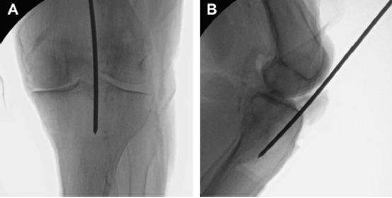
We need fluoroscopy to determine if the needle entry point is correct. It is very important to take a standard orthopantomogram of the knee intraoperatively, so how should it be taken?

Standard orthopantomogram-parallel line of the fibular head
The mechanical axis of the ortho-x-ray is made a straight line, and a parallel line of the mechanical axis is made at the lateral edge of the tibial plateau, which should bisect the fibular head on the ortho-x-ray. If one such x-ray is obtained, it proves to be taken correctly.

If the ortho-slice is not standard, for example, if the nail is fed from the standard feed point, when the external rotation position is taken, it will show that the feed point is outward, and the internal rotation position will show that the feed point is inward, which in turn will affect the surgical judgment.

On a standard lateral x-ray, the medial and lateral femoral condyles largely overlap and the medial and lateral tibial plateau largely overlap, and on the lateral view, the point of entry is located at the watershed between the plateau and the tibial stem.
IV. Content Summary
The standard tibial intramedullary nail entry point is located orthogonally at the medial edge of the lateral intercondylar spine of the tibia and laterally at the watershed between the tibial plateau and the tibial stem migration zone.
The safety zone at the entry point is very small, only 22.9±8.9 mm, and the needle can be inserted in this area without damaging the bony stop of the ACL and meniscal tissue.
Intraoperative standard orthopantomographs and lateral radiographs of the knee should be taken, which is the key to determine whether the needle entry point is correct or not.
Post time: Jan-02-2023


