


The meniscus is located between the medial and lateral femoral condyles and the medial and lateral tibial condyles and is composed of fibrocartilage with a certain degree of mobility, which can be moved along with the movement of the knee joint and plays an important stranded role in the straightening and stabilisation of the knee joint. When the knee joint moves suddenly and strongly, it is easy to cause meniscus injury and tear.
MRI is currently the best imaging tool for diagnosing meniscal injuries. The following is a case of meniscal tear provided by Dr Priyanka Prakash from the Department of Imaging, University of Pennsylvania, along with a summary of the classification and imaging of meniscal tears.
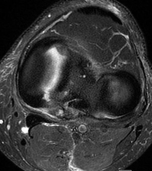
BASIC HISTORY: The patient had left knee pain for one week after a fall. The results of the MRI examination of the knee joint are as follows.
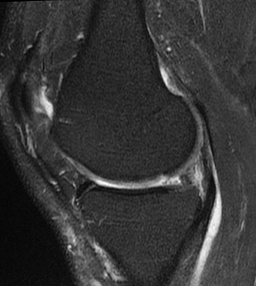
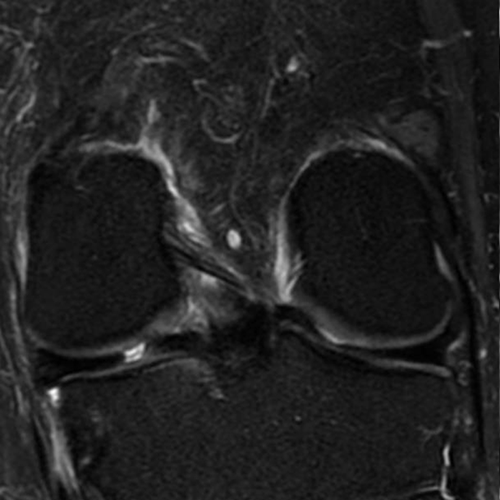
Imaging features: the posterior horn of the medial meniscus of the left knee is blunted, and the coronal image shows signs of a meniscal tear, which is also known as a radial tear of the meniscus.
Diagnosis: Radial tear of the posterior horn of the medial meniscus of the left knee.
Anatomy of the meniscus: On MRI sagittal images, the anterior and posterior corners of the meniscus are triangular, with the posterior corner larger than the anterior corner.
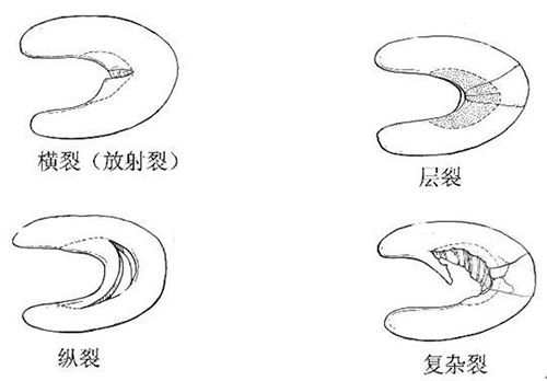
Types of meniscal tears in the knee
1. Radial tear: The direction of the tear is perpendicular to the long axis of the meniscus and extends laterally from the inner edge of the meniscus to its synovial margin, either as a complete or incomplete tear. The diagnosis is confirmed by the loss of the bow-tie shape of the meniscus in the coronal position and the blunting of the triangular tip of the meniscus in the sagittal position. 2. Horizontal tear: a horizontal tear.
2. Horizontal tear: A horizontally orientated tear that divides the meniscus into upper and lower parts and is best seen on MRI coronal images. This type of tear is usually associated with a meniscal cyst.
3. Longitudinal tear: The tear is orientated parallel to the long axis of the meniscus and divides the meniscus into inner and outer parts. This type of tear usually does not reach the medial edge of the meniscus.
4. Compound tear: a combination of the above three types of tears.
Magnetic resonance imaging is the imaging method of choice for meniscal tears, and for the diagnosis of a tear the following two criteria should be met
1. abnormal signals in the meniscus at least two consecutive levels to the articular surface;
2. abnormal morphology of the meniscus.
The unstable portion of the meniscus is usually removed arthroscopically.
Post time: Mar-18-2024


