


The acceptable criteria for the healing of humeral shaft fractures are an anterior-posterior angulation of less than 20°, lateral angulation of less than 30°, rotation of less than 15°, and shortening of less than 3cm. In recent years, with increasing demands for upper limb function and early recovery in daily life, surgical treatment of humeral shaft fractures has become more common. Mainstream methods include anterior, anterolateral, or posterior plating for internal fixation, as well as intramedullary nailing. Studies indicate that the nonunion rate for open reduction internal fixation of humeral fractures is approximately 4-13%, with iatrogenic radial nerve injury occurring in about 7% of cases.
To avoid iatrogenic radial nerve injury and reduce the nonunion rate of open reduction, domestic scholars in China have adopted the medial approach, using the MIPPO technique to fix humeral shaft fractures, and have achieved good results.

Surgical procedures
Step one: Positioning. The patient lies in a supine position, with the affected limb abducted 90 degrees and placed on a lateral operating table.
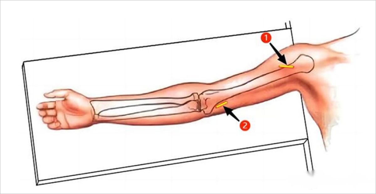
Step two: Surgical incision. In the conventional medial single-plate fixation (Kanghui) for patients, two longitudinal incisions of approximately 3cm each are made near the proximal and distal ends. The proximal incision serves as the entrance for the partial deltoid and pectoralis major approach, while the distal incision is located above the medial epicondyle of the humerus, between the biceps brachii and triceps brachii.
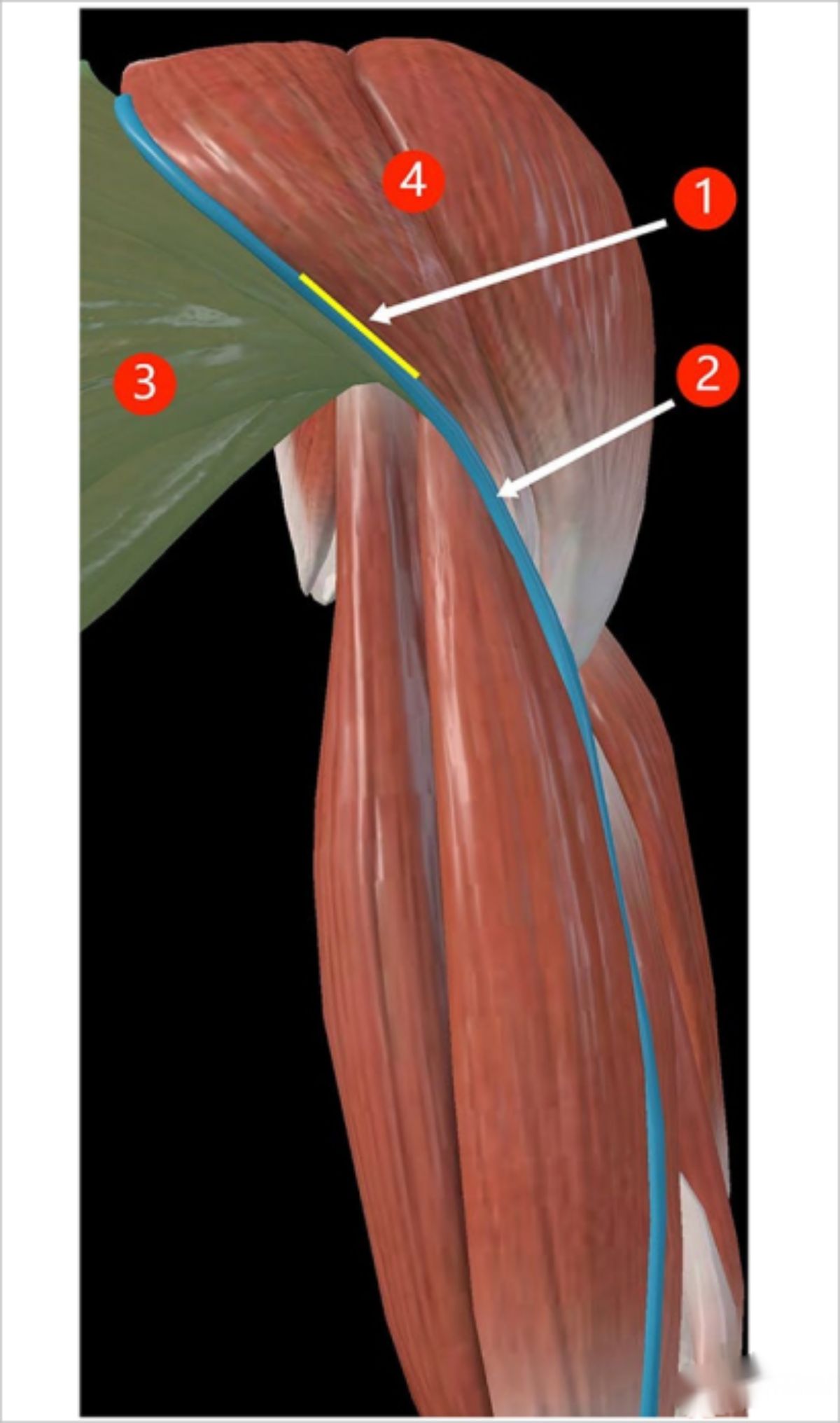
▲ Schematic diagram of the proximal incision.
①: Surgical incision; ②: Cephalic vein; ③: Pectoralis major; ④: Deltoid muscle.
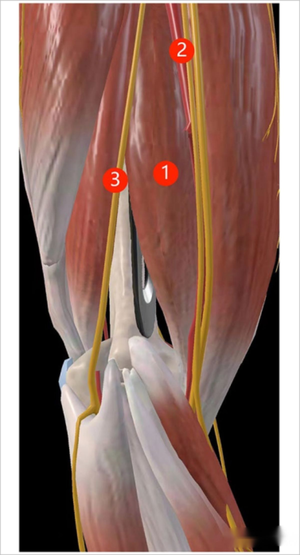
▲ Schematic diagram of the distal incision.
①: Median nerve; ②: Ulnar nerve; ③: Brachialis muscle; ④: Surgical incision.
Step three: Plate insertion and fixation. The plate is inserted through the proximal incision, snug against the bone surface, passing beneath the brachialis muscle. The plate is first secured to the proximal end of the humeral shaft fracture. Subsequently, with rotational traction on the upper limb, the fracture is closed and aligned. After satisfactory reduction under fluoroscopy, a standard screw is inserted through the distal incision to secure the plate against the bone surface. The locking screw is then tightened, completing the plate fixation.
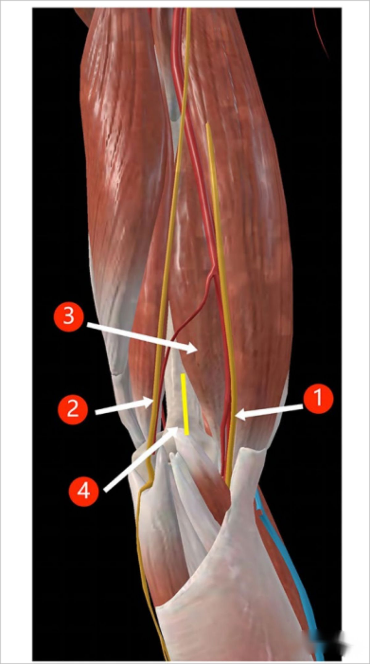
▲ Schematic diagram of the superior plate tunnel.
①: Brachialis muscle; ②: Biceps brachii muscle; ③: Medial vessels and nerves; ④: Pectoralis major.
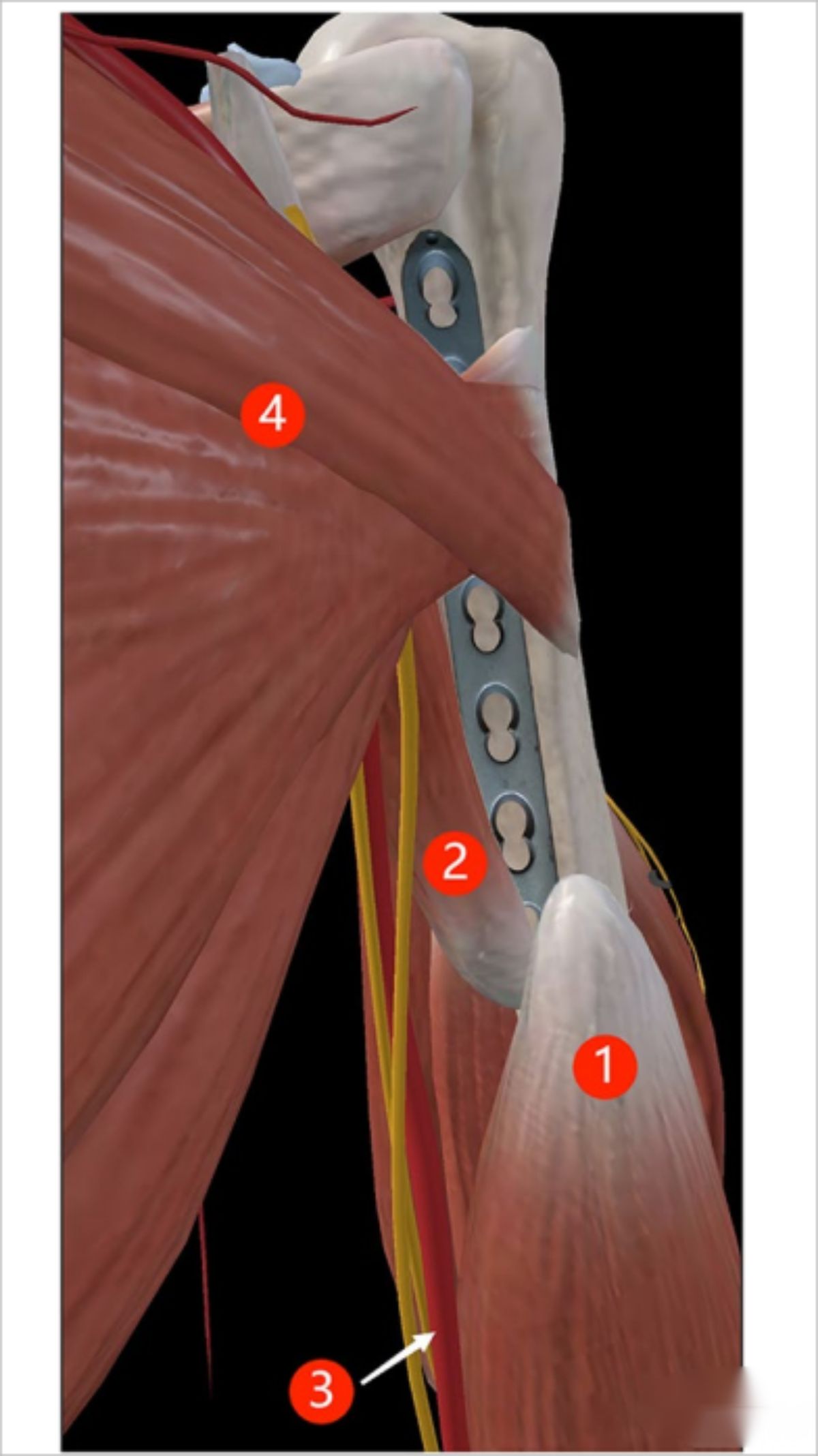
▲ Schematic diagram of the distal plate tunnel.
①: Brachialis muscle; ②: Median nerve; ③: Ulnar nerve.
Post time: Nov-10-2023


