


Femoral intertrochanteric fracture is the most common hip fracture in clinical practice and is one of the three most common fractures associated with osteoporosis in the elderly. Conservative treatment requires prolonged bed rest, posing high risks of pressure sores, pulmonary infections, pulmonary embolism, deep vein thrombosis, and other complications. The nursing difficulty is significant, and the recovery period is long, imposing a heavy burden on both society and families. Therefore, early surgical intervention, whenever tolerable, is crucial for achieving favorable functional outcomes in hip fractures.
Currently, the PFNA (proximal femoral nail antirotation system) internal fixation is considered the gold standard for surgical treatment of hip fractures. Achieving positive support during the reduction of hip fractures is crucial for allowing early functional exercise. Intraoperative fluoroscopy includes anteroposterior (AP) and lateral views to assess the reduction of the femoral anterior medial cortex. However, conflicts may arise between the two perspectives during surgery (i.e., positive in lateral view but not in anteroposterior view, or vice versa). In such cases, evaluating whether the reduction is acceptable and whether adjustment is needed poses a challenging problem for clinical practitioners. Scholars from domestic hospitals such as Oriental Hospital and Zhongshan Hospital have addressed this issue by analyzing the accuracy of assessing positive and negative support under anteroposterior and lateral views using postoperative three-dimensional CT scans as the standard.

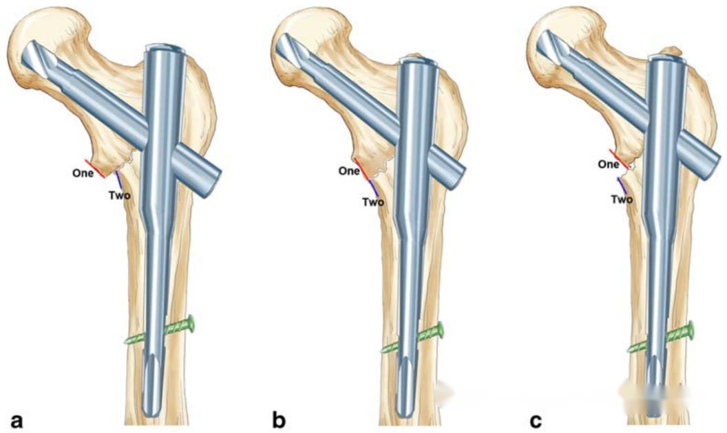
▲ The diagram illustrates positive support (a), neutral support (b), and negative support (c) patterns of hip fractures in the anteroposterior view.
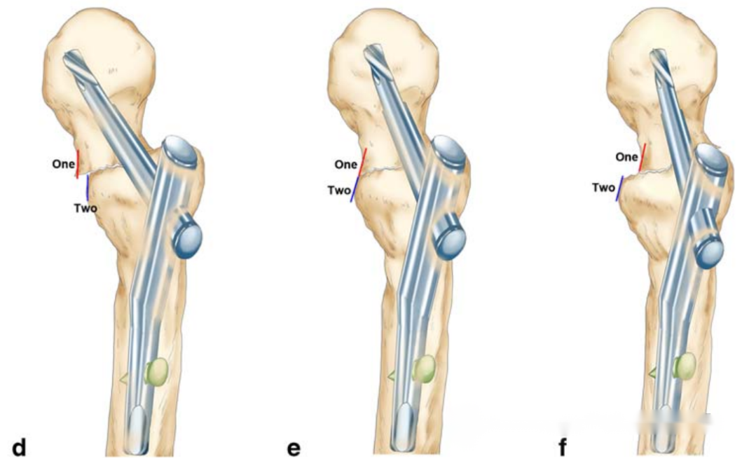
▲ The diagram illustrates positive support (d), neutral support (e), and negative support (f) patterns of hip fractures in the lateral view.
The article includes case data from 128 patients with hip fractures. Intraoperative anteroposterior and lateral images were separately provided to two doctors (one with less experience and one with more experience) to assess positive or non-positive support. After the initial assessment, a reevaluation was conducted after 2 months. Postoperative CT images were provided to an experienced professor, who determined whether the case was positive or non-positive, serving as the standard for evaluating the accuracy of the image assessments by the first two doctors. The main comparisons in the article are as follows:
(1)Are there statistically significant differences in the assessment results between the less experienced and more experienced doctors in the first and second assessments? Additionally, the article explores the intergroup consistency between less experienced and more experienced groups for both assessments and the intragroup consistency between the two assessments.
(2)Using CT as the gold standard reference, the article investigates which is more reliable for assessing the reduction quality: lateral or anteroposterior evaluation.
Research results
1. In the two rounds of assessments, with CT as the reference standard, there were no statistically significant differences in sensitivity, specificity, false positive rate, false negative rate, and other parameters related to the evaluation of reduction quality based on intraoperative X-rays between the two doctors with different levels of experience.
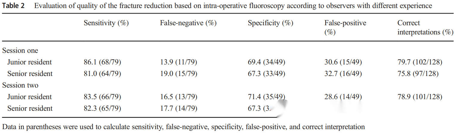
2.In the evaluation of reduction quality, taking the first assessment as an example:
- If there is agreement between anteroposterior and lateral assessments (both positive or both non-positive), the reliability in predicting reduction quality on CT is 100%.
- If there is disagreement between anteroposterior and lateral assessments, the reliability of lateral assessment criteria in predicting reduction quality on CT is higher.
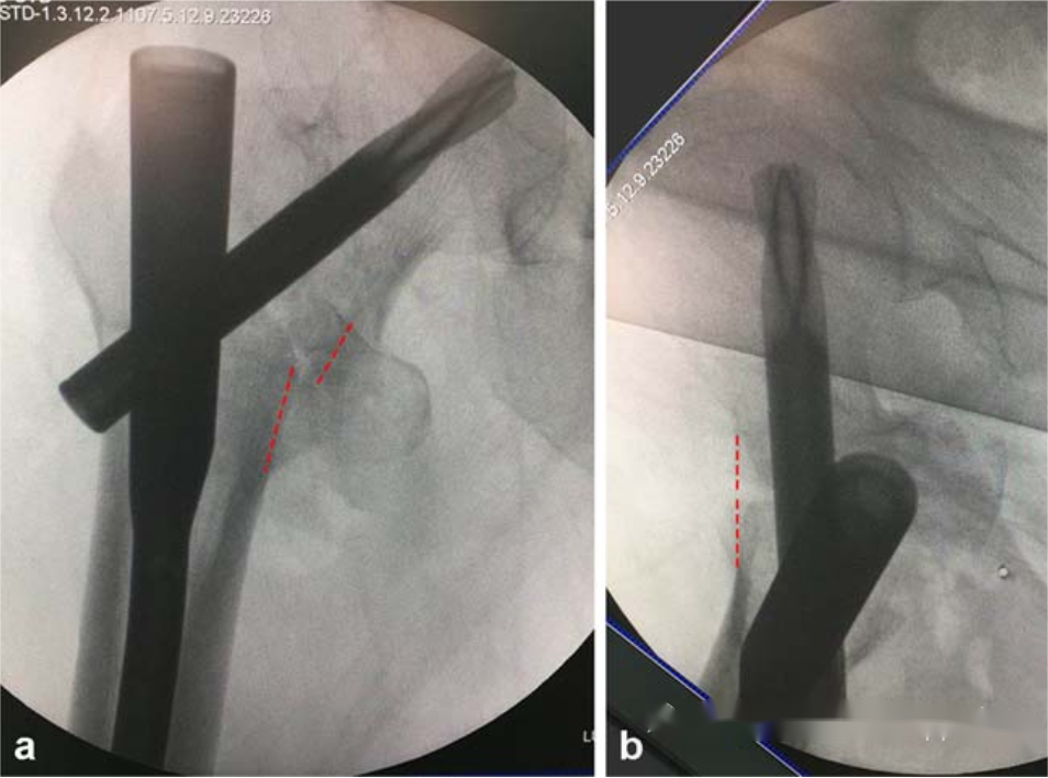
▲ The diagram illustrates a positive support shown in the anteroposterior view while appearing as non-positive in the lateral view. This indicates an inconsistency in the assessment results between the anteroposterior and lateral views.
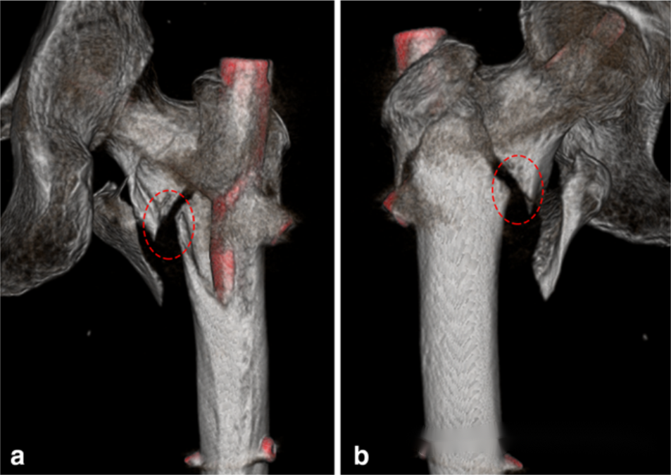
▲ Three-dimensional CT reconstruction provides multiple-angle observation images, serving as a standard for the assessment of reduction quality.
In the previous standards for reduction of intertrochanteric fractures, besides positive and negative support, there is also the concept of "neutral" support, implying anatomical reduction. However, due to issues related to fluoroscopy resolution and human eye discernibility, true "anatomical reduction" theoretically does not exist, and there are always slight deviations towards "positive" or "negative" reduction. The team led by Zhang Shimin at Yangpu Hospital in Shanghai published a paper (specific reference forgotten, would appreciate if someone can provide it) suggesting that achieving positive support in intertrochanteric fractures may result in better functional outcomes compared to anatomical reduction. Therefore, considering this study, efforts should be made during surgery to achieve positive support in intertrochanteric fractures, both in anteroposterior and lateral views.
Post time: Jan-19-2024


