


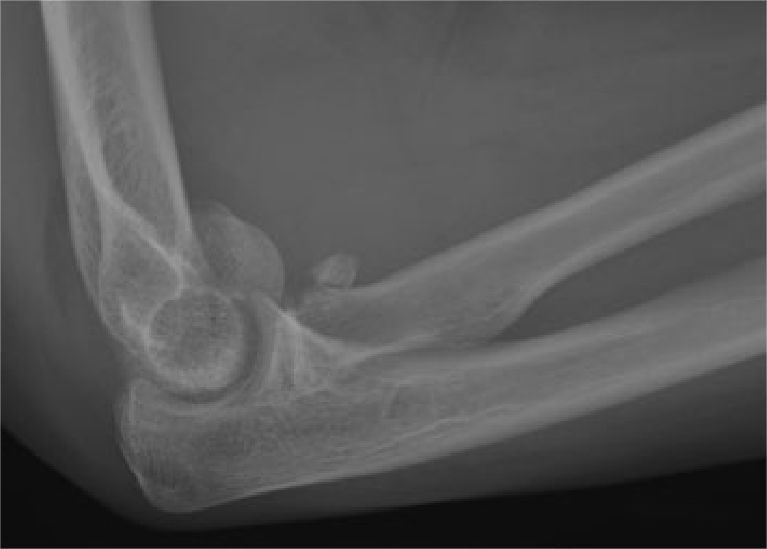
Fractures of the radial head and radial neck are common elbow joint fractures, often resulting from axial force or valgus stress. When the elbow joint is in an extended position, 60% of axial force on the forearm is transmitted proximally through the radial head. Following injury to the radial head or radial neck due to force, shearing forces can affect the capitulum of the humerus, potentially leading to bone and cartilage injuries.
In 2016, Claessen identified a specific type of injury where fractures of the radial head/neck were accompanied by bone/cartilage damage to the capitulum of the humerus. This condition was termed “kissing lesion,” with fractures that included this combination referred to as “kissing fractures.” In their report, they included 10 cases of kissing fractures and found that 9 cases had radial head fractures classified as Mason type II. This suggests that with Mason type II radial head fractures, there should be heightened awareness for potential accompanying fractures of the capitulum of the humerus.
In clinical practice, kissing fractures are highly prone to misdiagnosis, especially in cases where there is significant displacement of the radial head/neck fracture. This can lead to overlooking associated injuries to the capitulum of the humerus. To investigate the clinical characteristics and incidence of kissing fractures, foreign researchers conducted a statistical analysis on a larger sample size in 2022. The results are as follows:
The study included a total of 101 patients with radial head/neck fractures who were treated between 2017 and 2020. Based on whether they had an associated fracture of the capitulum of the humerus on the same side, the patients were divided into two groups: the capitulum group (Group I) and the non-capitulum group (Group II).

Furthermore, the radial head fractures were analyzed based on their anatomical location, which was divided into three regions. The first is the safe zone, the second is the anterior medial zone, and the third is the posterior medial zone.

The study results revealed the following findings:
- The higher the Mason classification of radial head fractures, the greater the risk of accompanying capitulum fractures. The probability of a Mason type I radial head fracture being associated with a capitulum fracture was 9.5% (6/63); for Mason type II, it was 25% (6/24); and for Mason type III, it was 41.7% (5/12).

- When radial head fractures extended to involve the radial neck, the risk of capitulum fractures decreased. The literature did not identify any isolated cases of radial neck fractures being accompanied by capitulum fractures.
- Based on the anatomical regions of radial head fractures, fractures located within the “safe zone” of the radial head had a higher risk of being associated with capitulum fractures.


▲ Mason classification of radial head fractures.


▲ A case of kissing fracture patient, where the radial head was fixed with a steel plate and screws, and the capitulum of the humerus was fixed using Bold screws.
Post time: Aug-31-2023


