Anterior screw fixation of the odontoid process preserves the rotational function of C1-2 and has been reported in the literature to have a fusion rate of 88% to 100%.
In 2014, Markus R et al published a tutorial on the surgical technique of anterior screw fixation for odontoid fractures in The Journal of Bone & Joint Surgery (Am). The article describes in detail the main points of the surgical technique, postoperative follow-up, indications and precautions in six steps.
The article emphasises that only type II fractures are amenable to direct anterior screw fixation and that single hollow screw fixation is preferred.
Step 1: Intraoperative positioning of the patient
1. Optimal anteroposterior and lateral radiographs must be taken for the operator’s reference.
2. The patient must be kept in the open-mouth position during surgery.
3. The fracture should be repositioned as much as possible before the start of surgery.
4. The cervical spine should be hyperextended as much as possible to obtain optimal exposure of the base of the odontoid process.
5. If hyperextension of the cervical spine is not possible – e.g., in hyperextension fractures with posterior displacement of the cephalad end of the odontoid process – then consideration may be given to translating the patient’s head in the opposite direction relative to his or her trunk.
6. immobilise the patient’s head in as stable a position as possible. The authors use the Mayfield head frame (shown in Figures 1 and 2).
Step 2: Surgical approach
A standard surgical approach is used to expose the anterior tracheal layer without damaging any important anatomical structures.
Step 3: Screw entry point
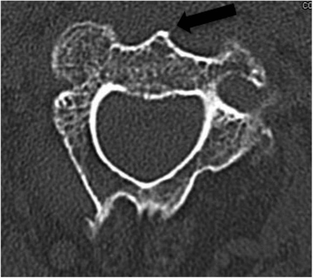
The optimal entry point is located at the anterior inferior margin of the base of the C2 vertebral body. Therefore, the anterior edge of the C2-C3 disc must be exposed. (as shown in Figures 3 and 4 below) Figure 3
The black arrow in Figure 4 shows that the anterior C2 spine is carefully observed during the preoperative reading of the axial CT film and must be used as an anatomical landmark for determining the point of needle insertion during surgery.
2. Confirm the point of entry under anteroposterior and lateral fluoroscopic views of the cervical spine. 3.
3. Slide the needle between the anterior superior edge of the C3 upper endplate and the C2 entry point to find the optimal screw entry point.
Step 4: Screw placement
1. A 1.8 mm diameter GROB needle is first inserted as a guide, with the needle orientated slightly behind the tip of the notochord. Subsequently, a 3.5 mm or 4 mm diameter hollow screw is inserted. The needle should always be slowly advanced cephalad under anteroposterior and lateral fluoroscopic monitoring.
2. Place the hollow drill in the direction of the guide pin under fluoroscopic monitoring and slowly advance it until it penetrates the fracture. The hollow drill should not penetrate the cortex of the cephalad side of the notochord so that the guide pin does not exit with the hollow drill.
3. Measure the length of the required hollow screw and verify it with the preoperative CT measurement to prevent errors. Note that the hollow screw needs to penetrate the cortical bone at the tip of the odontoid process (to facilitate the next step of fracture end compression).
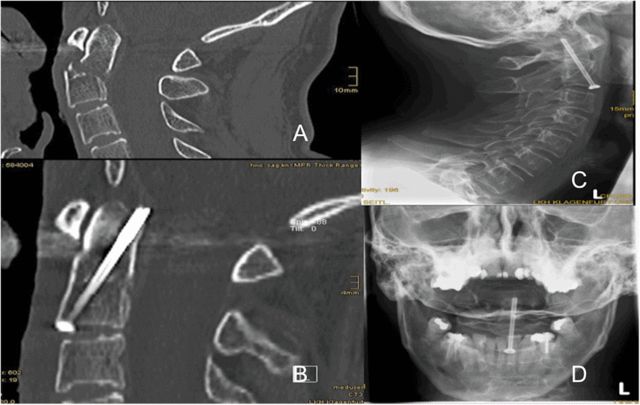
In most of the authors’ cases, a single hollow screw was used for fixation, as shown in Figure 5, which is centrally located at the base of the odontoid process facing cephalad, with the tip of the screw just penetrating the posterior cortical bone at the tip of the odontoid process. Why is a single screw recommended? The authors concluded that it would be difficult to find a suitable entry point at the base of the odontoid process if two separate screws were to be placed 5 mm from the midline of C2.
Figure 5 shows a hollow screw centrally located at the base of the odontoid process facing cephalad, with the tip of the screw just penetrating the cortex of the bone just behind the tip of the odontoid process.
But apart from the safety factor, do two screws increase postoperative stability?
A biomechanical study published in 2012 in the journal Clinical Orthopaedics and Related Research by Gang Feng et al. of the Royal College of Surgeons of the United Kingdom showed that one screw and two screws provide the same level of stabilisation in the fixation of odontoid fractures. Therefore, a single screw is sufficient.
4. When the position of the fracture and the guide pins are confirmed, the appropriate hollow screws are placed. The position of the screws and pins should be observed under fluoroscopy.
5. Care should be taken to ensure that the screwing device does not involve the surrounding soft tissues when performing any of the above operations. 6. Tighten the screws to apply pressure to the fracture space.
Step 5: Wound Closure
1. Flush the surgical area after completing screw placement.
2. Thorough haemostasis is essential to reduce postoperative complications such as haematoma compression of the trachea.
3. The incised cervical latissimus dorsi muscle must be closed in precise alignment or the aesthetics of the postoperative scar will be compromised.
4. Complete closure of the deep layers is not necessary.
5. Wound drainage is not a required option (authors usually do not place postoperative drains).
6. Intradermal sutures are recommended to minimise the impact on the patient’s appearance.
Step 6: Follow-up
1. Patients should continue to wear a rigid neck brace for 6 weeks postoperatively, unless nursing care requires it, and should be evaluated with periodic postoperative imaging.
2. Standard anteroposterior and lateral radiographs of the cervical spine should be reviewed at 2, 6, and 12 weeks and at 6 and 12 months after surgery. A CT scan was performed at 12 weeks after surgery.
Post time: Dec-07-2023


