


· Applied Anatomy
The entire length of the clavicle is subcutaneous and easy to visualize. The medial end or sternal end of the clavicle is coarse, with its articular surface facing inward and downward, forming the sternoclavicular joint with the clavicular notch of the sternal handle; the lateral end or acromion end is coarse and flat and wide, with its acromion articular surface ovoid and outward and downward, forming the acromioclavicular joint with the acromion. The clavicle is flat above and bluntly rounded in the middle of the anterior margin. There is a rough indentation of the costoclavicular ligament on the medial side below, where the costoclavicular ligament attaches. Lateral to the underside there is a conical node and oblique line with the conical ligament of the rostroclavicular ligament and the oblique ligament attachment, respectively.
· Indications
1. Clavicle fracture requiring incision and reduction internal fixation.
2. Chronic osteomyelitis or tuberculosis of clavicle requires dead bone removal.
3. Clavicle tumor requires resection.
· Body position
Supine position, with shoulders slightly elevated.
Steps
1. Make an incision along the S-shaped anatomy of the clavicle, and extend the incision along the upper edge of the clavicle to the inner and outer sides with the position of the lesion as a sign, and the site and length of the incision will be determined according to the lesion and the surgical requirements (Figure 7-1-1(1)).

Figure 7-1-1 Anterior Clavicular Manifestation Pathway
2. Incise the skin, subcutaneous tissue and deep fascia along the incision and free the skin flap up and down as appropriate (Figure 7-1-1(2)).
3. Incise the vastus cervicis muscle to the upper surface of the clavicle, the muscle is rich in blood vessels, pay attention to electrocoagulation. The periosteum is incised along the bony surface for subperiosteal dissection, with the sternocleidomastoid clavicle on the inner upper part, the pectoralis major clavicle on the inner lower part, the trapezius muscle on the outer upper part, and the deltoid muscle on the outer lower part. When stripping the posterior subclavian, the stripping should be performed tightly against the bone surface, and the control stripper should be steady so as not to damage the blood vessels, nerves, and pleura of the posterior clavicle (Figure 7-1-2). If it is proposed to apply the screw fixation of the plate, the soft tissues around the clavicle are first protected with the periosteal stripper, and the drill hole should be directed anteriorly downward, not posteriorly downward, so as not to injure the pleura and the subclavian vein.
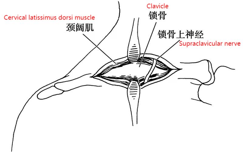 Figure 7-1-2 Exposing the Clavicle
Post time: Nov-21-2023


