


By CAH Medical | Sichuan, China
For buyers seeking low MOQs and high product variety, Multispecialty Suppliers offer low MOQ customization, end-to-end logistics solutions, and multi-category procurement, backed by their rich industry and service experience and strong understanding of emerging product trends.
I.What is the external fixation?
Common external fixators include plaster splints and small splints. Continuous traction (such as bone traction and skin traction) also has the function of reducing, braking, and correcting deformities, and is also a form of external fixation. In addition, external pinning fixation, which involves piercing the bone ends with steel needles and attaching external stents, is also a form of external fixation. It is mainly used for severe open fractures and severe soft tissue contusions, where external fixation is not possible and surgical internal fixation is difficult.
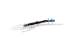
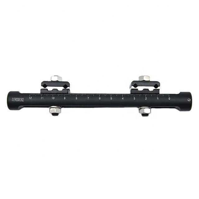
An external fixator is a device used to fix the affected limb externally. It holds the limb in the desired therapeutic position to facilitate the repair of fractures and other soft tissues. The purpose of an external fixator is to maintain a certain position to facilitate the repair of fractures and other soft tissues.
II.What is the procedure for external fixation?
External fixation is an orthopedic procedure used to treat bone conditions like fractures and dislocations. Here’s a concise summary:
Fracture Reduction:
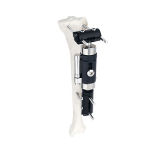
Reduction involves traction and manual rotation to correct pelvic displacements. For sacroiliac joint issues, the surgeon pushes the ilium toward the foot and spine. Bone traction is done by inserting a needle into the femoral condyle. In non-emergency cases, lower limb traction with 15-20 kg weight is used first. After reduction, a pelvic external fixator is applied, with 10 kg traction for 4-6 weeks. For anterior ring fractures without hemipelvic dislocation, only an external fixator is needed, not lower limb traction.
Needling:
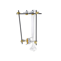
Identify bony landmarks like the iliac crest and anterior superior iliac spine. Kirschner wires are inserted percutaneously along the lateral iliac wall to determine the inclination of the iliac crest. Fixation pins are placed between the inner and outer iliac plates. Three 3mm wires are inserted in a parallel row along each iliac crest. A 5mm incision is made 2 cm posterior to the anterior superior iliac spine. Pins are inserted midway along the iliac crest into the medullary cavity, angled 15°-20° to the sagittal plane, pointing medially and downward, and secured approximately 5-6 cm deep.
Post time: Sep-16-2025


